28 diseases - Peripheral vascular disease

DNA test for genetic predisposition to peripheral vascular disease

Peripheral vascular disease (PVD) is a group of diseases characterized by narrowing of the lumen of the arteries supplying the limbs and internal organs. The danger of PVD lies in the deterioration of the function of the affected organ, with the risk of losing it altogether.

Warn yourself from diseases
Peripheral vascular disease

Damage to the blood vessels of the heart and brain is not classed as peripheral vascular disease: these pathologies belong to a separate group of diseases. In most cases, the morphological basis of the disease is atherosclerosis of the arteries with the formation of characteristic cholesterol plaques that obliterate (block) the lumen of the blood vessel. The site most commonly affected is the blood vessels of the legs. For this reason, PVD is often called obliterating atherosclerosis of lower limb arteries, or obliterating atherosclerosis of vessels in the lower extremities.

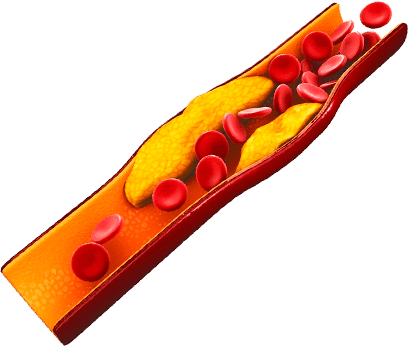
Atherosclerotic plaques form in the arteries when there is a metabolic dysfunction and a high concentration of low- and very low-density lipoproteins (LDL and VLDL) in the blood. These substances saturate the inner vessel wall, forming lipid spots where dense plaques then form, making the vascular lumen smaller, destroying the vessel wall and increasing the risk of thrombosis and accompanying thrombotic complications.

Peripheral vascular disease affects about 2.5% of the world population. In the USA, Europe and other developed countries, the morbidity rate in the 45-50 age group is 5.3%. Prevalence increases with age, the disease striking as many as 15% of the population by the age of 85. People in developing countries suffer the disease somewhat less often.

PVD is diagnosed late, when complications are already becoming apparent, in nearly half of all patients. If appropriate treatment is not given, nearly a third of patients die within five years of the first signs appearing, and indications for amputation occur in 50%.

Risk factors

The most important risk factors are:


Smoking;

Lipid exchange dysfunction;

Diabetes;

Improper diet with a high consumption of animal fats and simple carbohydrates (sugar and starchy foods);

High blood pressure;

Obesity;

Any atherosclerotic diseases (angina, cerebro-vascular disease etc);

Hereditary predisposition to atherosclerosis and peripheral vascular disease.

The statistics show that smoking is particularly significant: about 90% of all patients with diagnosed PVD are, or recently were, long-standing smokers. On average, smokers have a ten times higher risk of atherosclerotic damage to arteries in the leg than do non-smokers, and this correlation is twice as high for peripheral vascular disease than for coronary heart disease.
Genetic predisposition

Over half of patients with clinically expressed vascular atherosclerosis have an indication in their family history of relatives who have coronary heart disease, have had a stroke or another disease of atherosclerotic origin..

Genetic predisposition to peripheral vascular disease is related to certain features of lipid metabolism: in this case, lipid exchange does not ensure the optimal balance of different fractions of fat. Consequently, the level of cholesterol and low-density lipoproteins rises, and the concentration of high-density lipoproteins falls. This disproportion is what causes atherosclerotic plaques to form.

of high-density lipoproteins falls. This disproportion is what causes atherosclerotic plaques to form.

Another characteristic genetic feature of PVD patients is an atypical structure of the vascular wall, whose inner surface is particularly susceptible to damage under minimal excess loads (such as during a brief rise in blood pressure caused by stress). Microtrauma to the inner layer of the artery becomes the epicenter for the accumulation of atherogenic fats and the subsequent formation of the classic plaque.

Atherosclerosis of the lower limb arteries: symptoms and stages

Peripheral vascular diseases proceed in clearly delineated stages: beginning with inconspicuous manifestations, the blood supply dysfunction gradually progresses and, if left untreated, it may end with the death of tissues and the entire organ.

Atherosclerosis of vessels in the leg goes through three stages:

1

Asymptomatic atherosclerosis:
causes the patient no inconvenience; detectable only during a targeted examination;

2

Intermittent claudication:
characterized by periodic pain and numbness in the affected limb;

3

Critical limb ischemia:
the terminal stage of the process, with ulcer formation and possible progression to gangrene.


Asymptomatic atherosclerosis

The absence of any signs of dysfunction in the flow of blood along the lower limb vessels at the early stage is the result of the ability of the vascular network to compensate, and the adaptation of soft tissue to reduced blood supply and hypoxia.

Even so, the risk to such patients of atherosclerotic damage to the coronary and cerebral arteries is quite high.
Intermittent claudication

The full-blown stage of peripheral vascular disease is intermittent claudication. The symptoms indicate that the compensatory mechanisms are gradually becoming depleted and that significant tissue hypoxia is developing.

The following symptoms are characteristic of the disease:

  1. 1

    Pain: occurs suddenly after load (walking or running) and goes away after rest;

  2. 2

    Numbness or a tingling sensation under load;

  3. 3

    Low temperature of the affected limb (verified by comparing with the other limb);

  4. 4

    Changes in the function of the sebaceous and sweat glands: unilateral perspiration or, conversely, dry, peeling skin;

  5. 5

    Hair loss on the affected side;

  6. 6

    Reduced volume and fullness of soft tissue;

  7. 7

    Signs of osteoporosis (on an x-ray).

If untreated, the pathological process progresses with worsening symptoms: attacks of pain occur while at rest, and the patient has difficulty moving and needs third-party assistance.
Critical limb ischemia

In the disease’s last stage of development, the narrowing of the lumen reaches critical severity. The patient is troubled by constant pain that intensifies under load. Trophic, non-healing ulcers form on the skin of the affected limb; these easily become infected and suppurate. The limb feels almost cold to the touch, the pulse in its arteries feels weak, and blood pressure in the affected leg is low.

Without treatment, the gangrene and loss of the limb will follow.

Acute limb ischemia

A separate disease of the peripheral arteries is acute limb ischemia. This condition is caused by an embolism of one of the blood vessels and usually requires urgent surgical intervention, without which it is life-threatening. The embolus blocking the arterial lumen is usually a detached thrombus, or part of an atherosclerotic plaque from any location.

Acute limb ischemia manifests as sudden pain, tactile dysfunction and motor dysfunction. Without appropriate medical aid, irreversible changes appear within 3-4 hours: the soft tissues die and form areas of necrosis.

Take the DNA test
Check yourself and your family!

Every year, the health of millions of people is put at risk by late diagnosis and delayed treatment of peripheral vascular disease. Millions of dollars are spent on resolving the issues caused by the disease, but spending large sums of money is far from guaranteed to save the affected limb or to prevent serious complications.

Take care of yourself and your loved ones - take a DNA test for genetic predisposition to peripheral arterial disease!

Diagnosis

The following tests are used to make an exact diagnosis and to identify the early stages of peripheral arterial disease, including those that are symptom-free:


Examination of the limb
including measurement of arterial blood pressure and local skin surface temperature, and determination of pulse volume.

Calculation of the ABI
or Ankle-Brachial Index, defined as the ratio of the systolic arterial blood pressure (the higher of the two blood pressure figures) at the leg to the systolic pressure at the shoulder. With normal vascular patency, the blood pressure in the lower extremity is 10-15 mm of mercury higher than in the arm, giving an index greater than 1. Significant narrowing of the arterial lumen causes blood pressure to fall, giving an ABI of less than 1.

Calculation of the ABI under load:
enables detection of less pronounced arterial stenosis of the lower limbs.

Doppler ultrasound test:
analyses the blood flow through the lower limb vessels and detects volume deficiencies by the lowered blood pressure.

Contrast angiography:
carried out to visualize the condition of the arteries. An x-ray contrastive agent is injected into the blood vessel and a series of photographs are taken, showing the blood-filled arteries and areas of narrowing.

CT
or Computed Tomography: a highly informative technique for studying the arteries. Allows vascular stenosis to be observed and its severity assessed.

MRI
or Magnetic Resonance Imaging. This is a safe method of examination resulting in a three-dimensional representation of the blood vessels and soft tissues.

Laboratory testing of blood lipids
by fraction and of blood sugar levels (including analysis of glucose tolerance).

Atherosclerosis of lower limb blood vessels: treatment and prognosis

Treatment of atherosclerosis of the vessels of the lower extremities should be begun as early as possible. This will enable satisfactory blood supply to be maintained to the tissues, function to be supported at an adequate level and the development of complications to be prevented.

Effective treatment requires a complex approach and includes the following elements:

  1. 1

    Lifestyle adjustment:
    no smoking, dietary therapy, blood pressure control and physical activity;

  2. 2

    Treatment
    of diabetes, CHD and other atherosclerotic diseases;

  3. 3

    Therapeutic exercise:
    activates blood circulation and facilitates restoration of the vascular lumen;

  4. 4

    Drug therapy:
    hypolipemics to correct lipid exchange, antiplatelet agents and antiaggregants, drugs to relieve vascular spasm and reduce inflammation;

  1. 5

    Physiotherapy-based treatments:
    massage, electro-phonophoresis, cranial electrotherapy, UHV therapy, hydrotherapy;

  2. 6

    Revascularization:
    a host of methods enabling the flow of blood through the vessels to be restored. This can be done by inserting a shunt (bypass), balloon angioplasty (dilation of the lumen of the artery by means of a special balloon), reinforcement of the artery wall (stenting) and other relatively non-invasive forms of surgical treatment;

  3. 7

    Surgical methods in the late stages:
    these involve the removal of necrotized tissues. In life-threatening situations, the limb is amputated in order to preserve life and improve quality of life.

Prevention

Preventive measures are aimed at eliminating the risk factors:


Avoidance of harmful habits, especially smoking;

Correct diet;

Minimization of stress and overload;

Sufficient sleep;

Adequate physical activity;

Control of body weight and blood pressure;

Timely treatment of vascular, cardiac and endocrine diseases;

Regular preventive medical check-ups.

Your future in your hands!

The identification of a predisposition to atherosclerosis of the lower limbs can be used to make an individual prevention plan. A DNA test is the only way to determine the genetic risk of this dangerous disease. The test result will help with early diagnosis of insidious damage to the blood vessels.

Don’t wait for dangerous symptoms! Start prevention as early as possible: take a DNA test for genetic predisposition to peripheral vascular disease.